 EN
EN


Between 12.4 and 30% of children are born with spina bifida, also known as split spine. The prognosis and treatment depend on the stage of the disease. To date, it is impossible to fully cure it. However, there are many programs that can alleviate symptoms. With the right treatment, a happy and productive life with spina bifida is possible.
What is spina bifida?

Spina bifida is a congenital spinal cord defect that causes several vertebrae to split. As a result, the spinal cord is not protected by bone tissue. This is the organ that connects our brain to the rest of the body, and the disturbances in its work can cause many unpleasant consequences. In addition, the disturbances in the movement of cerebrospinal fluid can cause infections and brain damage.
The number and severity of symptoms depend on the stage of the disease. The most common are problems with urination and intestinal control, weakness and orthopedic diseases of the legs, low sensitivity, back pain, scoliosis and kyphosis, frequent fractures.
Bifida spina forms on the 28th day of pregnancy. There are medications that can reduce the risk of congenital neural tube pathologies. Usually, they are prescribed to women who have already had pregnancies with neurological abnormalities.
Spina bifida stages:
![]() Spina bifida occulta (closed type). This is the easiest stage. The spinal cord only slightly protrudes from the spine. This stage may be asymptomatic and only discovered by chance on the X-ray in childhood or adulthood. The appearance of the back may be normal, or it can be with slight swelling, discoloration, scars, hair growth, etc.
Spina bifida occulta (closed type). This is the easiest stage. The spinal cord only slightly protrudes from the spine. This stage may be asymptomatic and only discovered by chance on the X-ray in childhood or adulthood. The appearance of the back may be normal, or it can be with slight swelling, discoloration, scars, hair growth, etc.
Symptoms at this stage are not severe – gait disturbance, urinary issues, back pain, and scoliosis.
![]() Spina bifida meningocele. In this case, the spinal cord membranes are pushed outside the spine, forming a hernia. As a rule, the nerve endings remain protected by the membranes and the skin.
Spina bifida meningocele. In this case, the spinal cord membranes are pushed outside the spine, forming a hernia. As a rule, the nerve endings remain protected by the membranes and the skin.
The disease at this stage involves a risk of hydrocephaly and Chiari II malformation. These are neurological conditions caused by cerebrospinal fluid pressure on the brain. Without treatment, they can cause mental retardation, stunting and motor disorders.
![]() Myelomeningocele. At this stage, the spinal cord roots are displaced into the hernia. This form of the disease is the most dangerous and is associated with the greatest number of neurological complications. There is a high risk of hydrocephaly, paralysis, and meningitis.
Myelomeningocele. At this stage, the spinal cord roots are displaced into the hernia. This form of the disease is the most dangerous and is associated with the greatest number of neurological complications. There is a high risk of hydrocephaly, paralysis, and meningitis.
SPINA BIFIDA DIAGNOSTICS
Ultrasound
Medium and severe bifida spina can be diagnosed during pregnancy. The hernia can be seen on ultrasound scans starting from week 9. In addition, this pathology is indicated by defects in the legs, asymmetry of the ventricles of the brain and multi-hydration.
Still, spina bifida is difficult to diagnose by ultrasound.
Blood test
An increased amount of α-fetoprotein in the mother’s blood serum indicates fetal development defects. In combination with ultrasound, spina bifida is 80% likely to be detected in the third trimester.
MRI/CT
It allows doctors to see the extent of spinal damage and the location of the spinal cord. The process takes 40-90 minutes. It is important to lie still during the procedure, so children and those suffering from agoraphobia get anesthesia. In addition, in foreign clinics, there are open CT scanners, which are easier for anxious patients to tolerate.
DOPPLER ECHOCARDIOGRAPHY
Study of the blood supply to the heart with ultrasound. The results of this study are important to determine whether the patient can undergo the surgery.
NEUROSONOGRAPHY
In infants who have still have open skull fontanelle, ultrasound can detect hydrocephaly.
Electroneuromyography (ENMG)
The doctor checks the speed at which pulses pass through the neurons. In patients with spina bifida, the nerves of the lower limbs transmit the signal more slowly. The patient’s baseline is determined using this method, and a second study may reveal a decline in the condition.
Since most of the consequences caused by spina bifida affect the urinary system, urologist research is mandatory. Patients are prescribed urine tests, comprehensive urodynamic examination, ultrasound of the kidneys and bladder, cystoscopy (bladder probe studies), cystography and intravenous urology (X-ray examination with contrast agent).
SPINA BIFIDA SURGERY
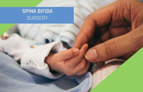
During the operation, the surgeon places the spinal cord tissue back into the spine and closes the opening with the help of muscles and skin. The earlier this procedure is done, the less damage will be done to the nerves.
После такой операции больной 4 дня находится в отделении интенсивной терапии. Еще 15 дней после этого за ним проводится стационарное наблюдение, и на протяжении 3 последующих месяцев проводятся обследования специалистами.
Bifida spine surgery is also possible during pregnancy. In this case, doctors perform an intrauterine operation. Depending on the period of pregnancy, the location of the fetus and the stage of the disease, it is possible to perform it through a fetoscopy or an incision in the mother’s abdomen.
Intrauterine surgery is associated with the best prognosis, but it can only be performed by experienced doctors who specialize in fetal surgery.
During the treatment of a hernia or hydrocephaly, doctors use a shunt to remove excess cerebrospinal fluid that puts pressure on the nerve ends. This shunt is changed from time to time due to the child’s growth or to avoid infection.
Untethering. Performed in order to treat of the fixed spinal cord. After the bifida spine surgery, the patient has a scar that fixes the spinal cord for a while and protects it from external influences. But as the child grows, the scar stretches, causing damage to the nerve tract. In this case, surgeons perform untethering surgery. Usually, children go through several such procedures as they grow up.
During this operation, the previous scar is cut to allow the surgeon access to the spinal cord. The doctor then separates the nerve pathways and the hernia from the scar tissue.
After this operation, the patient stays is in the intensive care unit for 4 days. For 15 more days they remain in the general ward for monitoring, and for the next 3 months, regular examinations are necessary.
The treatment of spina bifida also necessarily includes consultation with orthopedic surgeons and urologists. Depending on the extent of the patient’s impairment, they will select the safest and most effective procedures. Many children need catheterization and bladder enlargement.
CONSERVATIVE TREATMENT FOR SPINA BIFIDA
Children suffering from spina bifida need physiotherapy. Doctors create exercise programs to strengthen pelvic and leg muscles. Sometimes tires and canes are used to facilitate movement. Children with lower-limb paralysis may need a wheelchair or scooter.
In rehabilitation units, children can be taught basic household skills for self-care.
Treatment of spina bifida often involves taking antibiotics to avoid kidney or brain infection. Patients are also prescribed medication to facilitate urination and intestinal function.
SPECIFICS OF TREATING SPINA BIFIDA IN NEWBORNS
In the USA, in 66% of cases, the primary treatment for medium or severe spina bifida is given during pregnancy. If this is not possible, the child will be operated on within the first 48 hours of life. Cesarean section is used to avoid complications.
Surveys conducted in Ireland showed that 90% of children diagnosed with spina bifida face no difficulty in their daily lives.
Parents should know that due to frequent medical procedures, babies with spina bifida are prone to latex allergy. When caring for such children, you should use non-latex pacifiers, nipples, and gloves. Always inform health care workers if your child is allergic.
If the surgery has not yet been performed, the child is advised to sleep on the side or stomach rather than on the back. This will further protect the spinal cord.

Many children have decreased leg sensitivity. They may not notice the pain from physical damage. These children should not be allowed to run barefoot, and parents should avoid using hot-water heaters. In addition, kids may not feel that they are wearing too-tight shoes, which is bad for the development of their feet.
SPECIFICS OF TREATING SPINA BIFIDA IN ADULTS

For adults, unlike children, an operation is not necessary unless there is a sudden deterioration in symptoms. Often spina bifida only manifests at first in adult age. Usually, its exacerbation is caused by pregnancy, childbirth, physical activity, consequences of illness or surgery.
If this process has already begun, it will only get worse. The sooner the patient sees a doctor, the fewer negative consequences they will have to deal with.
The decision to carry out the operation is made taking into account the age of the patient and their general condition. Adults usually tolerate the intervention more easily. More procedures are also available for their treatment.
Although surgery is unable to repair damaged neurons, removing the hernia or cerebrospinal fluid pressure relieves many symptoms and improves the patients’ quality of life.
SYMPTOMS THAT REQUIRE IMMEDIATE TREATMENT
![]() Transparent yellow fluid leaking from the hernia or postoperative scar. This is cerebrospinal fluid, and through it, an infection can enter the spinal cord or brain. This condition can cause meningitis and irreversible brain damage.
Transparent yellow fluid leaking from the hernia or postoperative scar. This is cerebrospinal fluid, and through it, an infection can enter the spinal cord or brain. This condition can cause meningitis and irreversible brain damage.
![]() High fever for longer than three days without coughing or running a cold. This is most often a symptom of kidney inflammation.
High fever for longer than three days without coughing or running a cold. This is most often a symptom of kidney inflammation.
![]() Deterioration of symptoms: loss of leg function and sensitivity, spinal curvature, sagging feet, disorders of urination and bowel function, impaired vision, impaired swallowing and breathing, seizures, strabismus, pain, nausea, vomiting, learning difficulties.
Deterioration of symptoms: loss of leg function and sensitivity, spinal curvature, sagging feet, disorders of urination and bowel function, impaired vision, impaired swallowing and breathing, seizures, strabismus, pain, nausea, vomiting, learning difficulties.
IN WHICH FOREIGN CLINICS IS SPINA BIFIDA TREATED?
Among European clinics, you should pay attention to the Quironsalud network in Spain. Surgeries in these institutions are highly successful. One of the most outstanding children’s neurosurgeons in Madrid is Dr. José Hinojosa. He has extensive experience in the treatment of brain tumors, hydrocephaly and spina bifida.
Also, spina bifida is treated in the Czech Republic. Motol Hospital has a children’s neurosurgery department. It is one of the most technologically advanced medical institutions in Eastern Europe.
| Country | Cost |
|---|---|
| Turkey | from $10,000 |
| Spain | from €40,000 |
| The Chech Republic | from €18,000 |
| South Korea | from $17,000 |
Among the Turkish clinics, the most experienced doctors who deal with congenital spinal cord abnormalities can be found in Memorial and Liv clinics. In addition, Turkey’s rehabilitation centers have the most advanced physiotherapy programs for children with impaired mobility.
In South Korea, children’s neurosurgery is best performed at Soon Chun Hyang and Asan hospitals. This is the country that produces the best medical equipment, and Korean doctors are trained in the world’s most famous hospitals.
If you are looking for a hospital for spina bifida treatment, please leave your application on MedicGlobus website. Our coordinators will contact you to help choose the clinic and the surgeon, arrange your flight and treatment and support you throughout the process.

