EN
EN


Chiari malformation is an abnormal state, where brain tissue is shifted towards the spinal cord. It occurs in 1 person on 1,000. In most cases, the patient or their family do not notice any symptoms. In other cases, the disorder can cause risks to the patient’s health and life. To find out how to discover the malformation in time and treat it, read the article below.
What is Chiari malformation?
Chiari malformation is a structural defect that affects the cerebellum. It becomes displaced downwards one of both amygdalas through the foramen magnum (an opening in the bone of the skull nape).
This displacement has negative effects on the patient’s health. The damaged brain tissue fails at performing associated functions, and the cerebral fluid circulation gets blocked.
Congenital disorder forms during the mother’s pregnancy. It can be caused by genetic mutations, but also the mother’s nutrition.
There also is an acquired type of Chiari malformation. It develops on the later stages of development due to physical damage, illness or infection.
Congenital Chiari malformation is diagnosed in over 60% of the cases and acquired – only in 40%.

Chiari malformation symptoms
The main symptom of the disorder is a headache. It appears suddenly, after sneezing, coughing or due to stress. Other symptoms include:
 troubles with hearing;
tingle in the ears;
dizziness;
nausea and vomiting;
difficulty swallowing or speaking;
neck pain;
fine motor skills disorders;
muscle weakness;
limb numbness;
spinal curvature;
depression;
insomnia.
The exact symptoms that the patient suffers from depends on the damage to the neural tissue. Some people are not affected at all.
In newborns, Chiari malformation manifests with:
weak cries;
irritability during eating;
difficulty with swallowing;
excessive drooling;
frequent vomiting;
problems with neck movement;
breathing problems;
limb weakness;
problems with weight gain;
developmental delays.
Chiari malformation stages
| Type 1 | This type is found in most cases. The lower part of the cerebellum spreads to the skull opening. Childhood passes normally. First symptoms appear during the age of 25. Usually, the symptom is found by accident during examinations or testing for different disorders. |
| Type 2 | The disorder onsets in early childhood. The symptoms are more severe than in the first type. The complications can be life-threatening. Needs surgical intervention. |
| Type 3 | Very rare. A part of the cerebellum and/or spinal cord is pushed out through an opening in the back of the head or neck. Many such patients die soon after birth and those who survive to suffer from severe neurological defects. Requires early surgical intervention. |
| Type 4 | The rarest and severe type of disorder. The cerebellum cannot develop normally. Most children, born with this anomaly, do not survive. |
How is Chiari syndrome diagnosed?
Many patients live their lives without suffering from any of the described symptoms. The disease is often found during diagnostics for another disorder.
To confirm the diagnosis the doctors performs a medical examination, checks the patient’s memory, movement coordination, balance, reflexes, etc. This is done to assess the scale of the damage. Other necessary tests include:
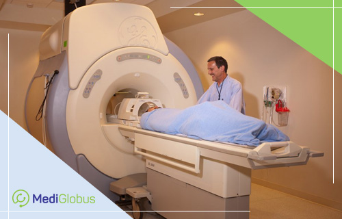
| CT | Computer tomography utilizes x-rays to make a visualization of the person’s inner body structure. Doctors usually use it to study the innate or acquired defects in bone integrity. |
| MRI | Similarly to the previous method, it is a way to visualize the inner structure of the patient’s body. However, instead of x-ray, it uses magnetic fields. This method is more effective in examining nerve tissue, visualizing its structure, position and assessing the damage. |
| Myelogram | Using a special needle, a surgeon injects contrast material into the patient’s liquid-filled spine segment. Due to the contrast interacting with the body tissues, it becomes possible to locate problematic areas of the spine, which will “light up” under x-rays. |
| VFSE | Video Fluoroscopic Swallowing Exam uses x-rays to observe the patient’s muscle movement during the process of swallowing objects of different, size, texture and consistency. This test is prescribed to patients who have trouble swallowing. |
| SSEP | Somatosensory Evoked Potential Test is a method of examining the spinal cord. Using special electrodes placed on legs or arms, the doctors send impulses up the spine and brain. Then, a different type of electrode is placed on the patient’s head or back. If any of the nerves that participate in the process suffer from compression, the test will show it. |
| BAER | Brainstem auditory evoked response is a test used to find anomalies in parts of the brain, involved in the hearing process. The specialist performing the test plays different sounds and measures the brainwave response of the patient. It is especially useful in diagnosing children, who cannot participate in usual auditory tests. |
How is Chiari malformation treated?

If the patient doesn’t suffer from any symptoms, there is no need to undergo invasive treatment. For this group of people, it would be sufficient to monitor their state by doing regular MRI scans.
Otherwise, the patient has to undergo surgery. The main treatment method for Chiari malformation is posterior fossa decompression. It is a surgical intervention during which the doctor creates space for the cerebellum by removing parts of bone tissue. Another benefit of the procedure is that it allows the cerebral fluid to renown its flow.
Decompression surgery is most effective when performed during pregnancy. This lowers the chances of hydrocephalus and other syndromes that cause nerve damage.
Some patients may need to undergo cervical spine fusion. Many also require the surgery to be repeated several times.
In which clinics is Chiari malformation treated?
Teknon Clinic in Spain
Soon Chun Hyang Hospital in South Korea
Helios Clinic in Germany
Assuta Medical Center in Israel
Navarra University Hospital in Spain
Liv Medical Network in Turkey
Resume
Chiari malformation is a structural defect, which causes cerebellum to be shifted towards the spinal cord. In some patients, this symptom doesn’t cause any symptom, but in other, it can severely affect their life and cause a lethal outcome.
The tests used to diagnose Chiari malformation include CT, MRI, myelogram, brainstem auditory evoked response, somatosensory evoked potential test, and fluorography.
The main method of treatment is posterior fossa decompression surgery. This procedure is most effective if performed during pregnancy.
Chiari malformation is treated in TOP hospitals of Turkey, Spain, Germany, Israel, the Czech Republic and South Korea.
To arrange a trip abroad to diagnose and treat Chiari malformation, contact the coordinating physicians of the MedicGlobus international platform. Leave a request and our specialists will reply as soon as possible.